What Does Eom Stand for in Medical Terms
What Does Eom Stand for in Medical Terms
EOM
Farlex Partner Medical Dictionary © Farlex 2012
EOM
Extraocular movements, see there.McGraw-Hill Concise Dictionary of Modern Medicine. © 2002 by The McGraw-Hill Companies, Inc.
muscle
(mus'el ) [L. musculus, diminutive of mus, mouse] 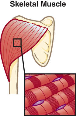
![]()
MORPHOLOGICAL FORMS OF MUSCLE
![]()
MORPHOLOGICAL FORMS OF MUSCLE
![]()
MORPHOLOGICAL FORMS OF MUSCLE

![]()
MORPHOLOGICAL FORMS OF MUSCLE
![]()
MORPHOLOGICAL FORMS OF MUSCLE
![]()
MORPHOLOGICAL FORMS OF MUSCLE
A type of tissue composed of contractile cells. Each muscle cell is filled with parallel actin and myosin filaments. When activated by an internal release of calcium, the filaments use the energy in ATP to crawl along each other in opposite directions. This movement shortens the length of the cell, which then contracts.
The three general classes of muscle cells (myocytes) are skeletal (striated), cardiac (striated), and smooth; most of the muscle in humans is skeletal. A typical muscle has a central portion called the belly and two or more attachment ends with tendons; the more stationary of the attachments is called the muscle's origin, while the more movable attachment is called the muscle's insertion. See: illustration
![]()
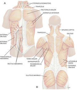
MUSCLES OF THE TRUNK
abdominal muscles
The abdominal muscles are made up of the cremaster, external abdominal oblique, iliacus, psoas major, pyramidalis, quadratus lumborum, rectus abdominis, and transversus abdominis muscles.
illustrationabducens muscle
, abducens oculiLateral rectus muscle, one of the extraocular muscles. Nerve: cranial nerve (CN VI). In clinical practice, referred to as the lateral rectus muscle.
abductor muscle
A muscle that on contraction draws a part away from the median plane of the body or the axial line of an extremity.
See: adductor muscleabductor digiti minimi muscle
Hand muscle. Origin: pisiform bone of wrist. Insertion: base of proximal phalanx of digit 5. Nerve: ulnar (C8-T1). Action: abducts digit 5.
abductor pollicis brevis muscle
Hand muscle. Origin: flexor retinaculum of wrist, scaphoid and trapezium bones. Insertion: lateral base of proximal phalanx of thumb. Nerve: median (C8-T1). Action: abducts thumb, aides in opposition with digit 5.
See: armfor illus. (Muscles of the Arm)adductor muscle
A muscle that draws toward the midline.
See: abductor muscleadductor brevis muscle
A muscle of the medial thigh originating on the ramus of the pubis and inserted in the linea aspera of the femur. It adducts, flexes, and medially rotates the thigh and is controlled by the obturator nerve.
adductor longus muscle
Hip and thigh muscle. Origin: front of pubis (below crest). Insertion: linea aspera of femur. Nerve: obturator (L2-L4). Action: adducts, flexes, and rotates thigh medially.
See: leg for illus. (Muscles of the leg)adductor magnus muscle
Hip and thigh muscle. Origin: inferior ramus of pubis, ramus of ischium, ischial tuberosity. Insertion: linea aspera and adductor tubercle of femur. Nerve: obturator and sciatic (L2-L4). Action: adducts, flexes, and rotates thigh medially.
See: leg for illus. (Muscles of the leg)adductor pollicis muscle
Hand muscle. Origin: capitate bone of wrist and metacarpals 2-3. Insertion: proximal phalanx of thumb and medial sesamoid bone. Nerve: ulnar (C8-T1. Action: adducts thumb, aides in opposition with digit 5.
agonist muscle
Controlled movements involve two opposing muscles: the agonist muscle produces the main action, while the antagonist muscle produces the opposite action to a lesser degree. The balance between agonist and antagonist muscles allows precise control of the final action.
Synonym: antagonist muscle See: PNF Stretching Techniquesanconeus muscle
A short muscle along the back of and outside the elbow. It originates from the lateral epicondyle of the humerus, crosses the back of the elbow joint on the same side, attaches to the lateral surface of the olecranon process and the adjacent surface of the ulna. It extends the forearm and abducts the elbow as the forearm pronates. It is innervated by the radial nerve (C7, C8, T1).
antagonist muscle
Agonist muscle.antigravity muscles
Muscles that pull against gravity to maintain normal posture.
Synonym: postural musclesappendicular muscle
One of the skeletal muscles of the limbs.
arrector pili muscle
Arrector pili.arm muscle
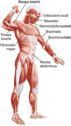
Arm: biceps brachii, brachialis, coracobrachialis, and triceps muscles. Forearm, anterior: flexor carpi radialis, flexor carpi ulnaris, flexor digitorum profundus, flexor digitorum superficialis, flexor pollicis longus, and pronator quadratus muscles. Forearm, posterior: abductor pollicis longus, anconeus, brachioradialis, extensor carpi radialis brevis, extensor carpi radialis longus, extensor carpi ulnaris, extensor digitorum, extensor digitorum minimi, extensor indicis, extensor pollicis brevis, extensor pollicis longus, and supinator muscles.
See: arm for illus. (Muscles of the Arm)articular muscle
A muscle attached to the capsule of a joint.
arytenoid muscle
The oblique or the transverse arytenoid -- laryngeal muscles. Origins: arytenoid cartilage. Insertions: contralateral arytenoid cartilage. Nerve: recurrent laryngeal and superior laryngeal of the vagus (CN X). Action: closes laryngeal inlet by bringing arytenoid cartilages toward each other.
auditory muscles
The tensor tympani and stapedius muscles.
axial muscle
A skeletal muscle that moves or stabilizes the head or the trunk.
back muscle
Superficial: latissimus dorsi and trapezius muscles. Middle layer: levator scapulae, rhomboid major, and rhomboid minor muscles. Deep layer: erector spinae and splenius. Deepest layer: interspinalis, intertransverse, multifidus, rotatores, semispinalis, and spinalis capitis.
biceps brachii muscle
Arm muscle. Origin: supraglenoid tubercle, coracoid process of scapula. Insertion: tuberosity of radius, posterior border of ulna (via bicipital aponeurosis). Nerve: musculocutaneous (C5-C6). Action: flexes forearm, supinates hand.
See: arm for illus. (Muscles of the Arm)biceps femoris muscle
Leg muscle. Origin: ischial tuberosity, linea aspera and second supracondylar ridge of femur. Insertion: lateral condyle of tibia, head of fibula. Nerve: sciatic (L5-S2). Action: flexes leg, rotates leg laterally, extends thigh.
See: leg for illus. (Muscles of the leg)bipennate muscle
A muscle in which the fibers converge from both sides to a central tendon.
illustrationbrachialis muscle
Arm muscle. Origin: anterior surface of lower (distal) humerus. Insertion: coronoid process of ulna. Nerve: musculocutaneous and radial (C5-C7). Action: flexes forearm.
See: arm for illus. (Muscles of the Arm)brachioradialis muscle
Arm muscle. Origin: lateral supracondylar ridge of distal humerus. Insertion: distal end of radius. Nerve: radial (C5-C7). Action: flexes forearm.
See: arm for illus. (Muscles of the Arm)buccinator muscle
Facial muscle. Origin: pterygomandibular raphe and alveolar processes of jaws. Insertion: orbicularis oris muscle at angle of mouth. Nerve: facial (CN VII). Action: compresses check against teeth, retracts angle of mouth.
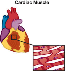
cardiac muscle
A tissue composed of mitochondrion-filled muscle cells that also contain neatly packed actin and myosin filaments; the filaments are arranged in cylindrical bundles called myofibrils. In each cell, the myofibrils are all aligned in the same direction and are parceled into longitudinal blocks (called sarcomeres) of similar lengths. Under the microscope, the ends of the blocks appear as lines, making cardiac muscle cells appear to have regularly arranged striations. In the muscle tissue, the cardiac muscle cells are connected in branching networks.
Cardiac muscle is innervated by both sympathetic and parasympathetic autonomic motor axons. In addition, cardiac muscle: is stimulated by blood—borne molecules, can conduct electrical impulses from cell to cell, and can independently generate rhythmical contractions. Cardiac muscle, which is found only in the heart, cannot be controlled consciously.
chest wall muscle
Pectoralis major, pectoralis minor, serratus anterior, subclavius, subscapularis, or teres major muscle.
chewing muscle
Mastication muscle.ciliary muscle
Internal eye muscle. Origin: edges of sclera. Insertion: ciliary process of lens. Nerve: oculomotor (CN III). Action: allows lens to become more curved to focus on near objects.
constrictor muscle of pharynx
A muscle that constricts the pharynx; it is important for swallowing.
core muscle
One of the major muscles that stabilizes and controls the pressure inside the trunk; these are the pelvic floor, abdominal wall, back, and diaphragm muscles.
corrugator muscle
Facial muscle. Origin: medial part of supraorbital margin. Insertion: skin above middle of eyebrow. Nerve: facial (CN VII). Action: pulls eyebrows toward midline and downward.
Synonym: Corrugator superciliicremaster muscle
Spermatic cord muscle. Origin: inguinal ligament and pubic tubercle. Insertion: cremasteric fascia covering spermatic cord. Nerve: genitofemoral (L1-L2). Action: elevates testis in males.
See: penis for illus.cricoarytenoid muscle
The lateral or the posterior cricoarytenoid -- laryngeal muscles. Origin: cricoid cartilage. Insertion: muscular process of arytenoid cartilage. Nerve: recurrent laryngeal of the vagus (CN X). Action: rotates arytenoid cartilages for vocalizations.
cricothyroid muscle
Laryngeal muscle. Origin: cricoid cartilage. Insertion: lower edges of thyroid cartilage. Nerve: superior laryngeal of the vagus (CN X). Action: tenses (stretches) vocal cords
See: thyroidfor illus.deep neck muscle
One of the various neck muscles that surround the vertebral column and base of the skull and which are contained in the prevertebral cylinder of deep cervical fascia. All these muscles are innervated by cervical spinal nerves, and most of these muscles act primarily to move and stabilize the head.
deltoid muscle
Shoulder muscle. Origin: a bony ellipse from the lateral third of the clavicle over the acromial process and along the spine of the scapula. Insertion: deltoid tuberosity on the lateral shaft of the humerus. Nerve: axillary (C5-C6). Action: abducts arm.
See: arm for illus. (Muscles of the Arm)detrusor muscle
The three-layered muscular wall of the urinary bladder. Nerve: primarily parasympathetic (S2-S4), secondarily sympathetic (T11-L2). Action: empties bladder.
diaphragm muscle
Origin: internal surfaces of lower six ribs, xiphoid process, vertebral bodies L1-L3. Insertion: central tendon (of diaphragm). Nerve: phrenic, lower six intercostals. Action: inflates lungs
digastric muscle
Neck muscle with two bellies. Origin: anterior belly attaches to the digastric fossa in mandible at base of anterior midline, posterior belly attaches to mastoid process. Insertion: tendon connecting both bellies in a loop of fascia that is attached to hyoid bone. Nerve: anterior belly -- trigeminal (CN V), posterior belly -- facial (CN VII). Action: lowers mandible and raises hyoid bone.
See: neck for illus.erector spinae muscle
Three adjacent vertical bands of deep back muscles -- the iliocostalis, longissimus, and spinalis muscles. Origins: a wide tendon running along the iliac crest to the sacrum, the lower lumbar and sacral spinous processes. Insertions: along the back in the angles of the lower ribs, transverse processes of the thoracic and cervical vertebrae. Nerves: dorsal rami of the spinal nerves. Actions: extends (bends backward) the vertebral column and neck, twists the back.
extensor carpi ulnaris muscle
Forearm muscle. Origin: lateral epicondyle of humerus, proximal edge of ulna. Insertion: proximal end of fifth metacarpal. Nerve: radial (C7-C8). Action: adducts hand, extends wrist.
See: arm for illus. (Muscles of the Arm)extensor digitorum muscle
Forearm muscle. Origin: lateral epicondyle of humerus. Insertion: common extensor tendon of fingers. Nerve: radial (C7-C8). Action: extends fingers and wrist.
See: arm for illus. (Muscles of the Arm)extensor digitorum brevis muscle
Foot muscle. Origin: dorsolateral surface of calcaneus. Insertion: extensor tendons of toes. Nerve: deep peroneal (S1-S2). Action: extends toes.
See: leg for illus. (Muscles of the leg)extensor digitorum longus muscle
Foot muscle. Origin: lateral condyle of tibia, upper three-fourths of fibula. Insertion: extensor tendons of toes 2-5. Nerve: deep peroneal (L5-S1). Action: extends toes, dorsiflexes foot.
See: leg for illus. (Muscles of the leg)extensor hallucis longus muscle
Foot muscle. Origin: middle of fibula. Insertion: base of proximal phalanx of big toe. Nerve: deep peroneal (S1-S2). Action: dorsiflexes big toe.
See: leg for illus. (Muscles of the leg)external intercostal muscles
The outer layer of muscles between the ribs, originating on the lower margin of each rib and inserted on the upper margin of the next rib. During inspiration, they draw adjacent ribs together, pulling them upward and outward, and increasing the volume of the chest cavity. They are controlled by the intercostal nerves.
external oblique muscle
Abdominal wall muscle. Origin: lower costal margin. Insertion: anterior half of iliac crest, rectus sheath, inguinal ligament. Nerve: intercostals 8-12, iliohypogastric, ilioinguinal (L1). Action: tenses and compresses abdomen, flexes and laterally rotates spine, lowers rib cage.
external pterygoid muscle
Lateral pterygoid muscle.extraocular muscle
Abbreviation: EOMSix muscles that attach outside the eyeball and that move the eye in its socket. The EOM are: the inferior and superior oblique muscles, and the lateral, medial, inferior, and superior rectus muscles.
See: extraocular for illus.extrinsic muscle
Abbreviation: EMThe muscles outside an organ that control its position, such as the EM of the eye or tongue.
muscles of facial expression
Thin muscles that insert into the skin of the face; all are innervated by the facial nerve (CN VII). Scalp: frontalis and occipitalis muscles. Ear: anterior, posterior, and superior auricular muscles. Eye: orbicularis oculi. Nose: depressor septi, nasalis, and procerus muscles. Mouth: buccinator, depressor anguli oris, depressor labii inferioris, levator anguli oris, levator labii superioris, mentalis, orbicularis oris, risorius, and zygomaticus muscle. Neck: platysma.
See: face and headfor illus.muscles of facial expression
Facial muscles.
fibularis muscles
The newer name for the peroneus muscles.
fibularis longus muscle
Peroneus longus muscle.fixation muscle
A muscle that steadies a part so that more precise movements in a related structure may be accomplished.
flexor carpi radialis muscle
Forearm muscle. Origin: medial epicondyle of humerus. Insertion: bases of second and third metacarpals. Nerve: median (C6-C7). Action: abducts hand, flexes wrist.
See: Arm, muscles of the arm (illus.)flexor carpi ulnaris muscle
Forearm muscle. Origin: medial epicondyle of humerus, medial side of olecranon, proximal posterior edge of ulna. Insertion: pisiform, hamate, and base of fifth metacarpal. Nerve: ulnar (C7-C8). Action: adducts hand, flexes wrist.
flexor digitorum longus muscle
Foot muscle. Origin: posterior surface of middle tibia. Insertion: distal phalanges of toes 2-5. Nerve: tibial (S2-S3). Action: flexes toes 2-5, plantarflexes foot.
See: leg for illus. (Muscles of the leg)flexor digitorum profundus muscle
Forearm muscle. Origin: proximal three-fourths of ulna. Insertion: distal phalanges of fingers (digits 2-5). Nerve: ulnar, median (C8-T1). Action: flexes distal finger joints, aids in wrist flexion.
flexor digitorum superficialis muscle
Forearm muscle. Origin: medial epicondyle of humerus, coronoid process of ulna. Insertion: middle phalanges of fingers (digits 2-5). Nerve: median (C7-T1). Action: flexes fingers and wrist.
See: arm for illus. (Muscles of the Arm)flexor hallucis longus muscle
Foot muscle. Origin: distal two-thirds of posterior tibia. Insertion: plantar side of distal phalanx of big toe. Nerve: tibial (S2-S3). Action: flexes big toe, plantarflexes foot.
flexor pollicis brevis muscle
A muscle of the hand originating on the flexor retinaculum and trapezium, trapezoid, and capitate and inserted on the lateral side of the base of the first phalanx of the thumb. It flexes the thumb at both the carpometacarpal joint and the metacarpophalangeal joint and is controlled by the median and the ulnar nerves.
flexor pollicis longus muscle
Forearm muscle. Origin: coronoid process of ulna, anterior surface of radius. Insertion: distal phalanx of thumb. Nerve: median (C8-T1). Action: flexes thumb.
See: arm for illus. (Muscles of the Arm)foot muscles
Dorsal: dorsal interosseous, extensor digitorum brevis, extensor digitorum longus, extensor hallucis longus, and tibialis anterior muscles. Plantar: abductor digiti minimi, abductor hallucis, adductor hallucis, flexor digitorum brevis, flexor digiti minimi brevis, flexor hallucis brevis, lumbrical, plantar interosseous, and quadratus plantae muscles.
See: leg for illus. (Muscles of the leg)frontalis muscle
Front half of occipitofrontalis muscle – a facial muscle. Origin: epicranial (scalp) aponeurosis. Insertion: skin of eyebrows, root of nose. Nerve: facial (CN VII). Action: elevates eyebrows, wrinkles forehead.
See: face and head for illus.fusiform muscle
gastrocnemius muscle
Leg muscle. Origin: medial condyle of femur, lateral condyle of femur. Insertion: calcaneus (via Achilles tendon). Nerve: tibial (S1-S2). Action: plantarflexes foot, flexes knee.
See: leg for illus. (Muscles of the leg)gemellus muscle
Either of the two muscles that attach to the medial surface of the greater trochanter of the femur (the trochanteric fossa) where they mesh with the tendon of the obturator internus muscle. The superior gemellus muscle arises from the ischial spine and is innervated by the nerve to the obturator internus; the inferior arises from the ischial tuberosity and is innervated by the femoral nerve. Both muscles hold the head of the femur in the acetabulum, rotate (laterally) the thigh in extension, and abduct the thigh when it is flexed.
genioglossus muscle
Tongue muscle. Origin: genial tubercle on inside of mandibular symphysis. Insertion: ventral tongue, hyoid bone. Nerve: hypoglossal (CN XII). Action: protrudes and depresses tongue.
gluteus maximus muscle
Thigh muscle. Origin: upper outer edge of ilium and sacrum. Insertion: iliotibial tract of fascia lata, gluteal tuberosity of femur. Nerve: inferior gluteal (L5-S2). Action: extends, abducts, and laterally rotates thigh.
gluteus medius muscle
Thigh muscle. Origin: lower half of ilium. Insertion: proximal medial tibia. Nerve: obturator (L2-L3). Action: adducts, flexes, and medially rotates thigh.
gracilis muscle
Thigh muscle. Origin: lower half of pubis. Insertion: proximal medial tibia. Nerve: obturator (L2-L3). Action: adducts, flexes, and medially rotates thigh.
See: leg for illus. (Muscles of the leg)hamstring muscles
Posterior thigh muscles that originate on the ischial tuberosity and act across both the hip and knee joints; they are the biceps femoris, gracilis, sartorius, semitendinosus, and semimembranosus muscles.
hand muscles
Abductor digiti minimi, abductor pollicis brevis, adductor pollicis, dorsal interosseous, flexor digiti minimi, flexor pollicis brevis, lumbrical, opponens digiti minimi, opponens pollicis, palmaris brevis, and palmar interosseous muscles.
Hilton muscle
See: Hilton, Johnhyoglossus muscle
A sheet of muscle extending up from the hyoid bone to the ipsilateral base and sides of the tongue. It depresses the sides of the tongue and is innervated by cranial nerve XII (hypoglossal nerve).
iliacus muscle
Thigh muscle. Origin: iliac fossa. Insertion: lesser trochanter of femur, psoas major tendon. Nerve: femoral (L2-L3). Action: flexes thigh.
iliopsoas muscle
The iliacus and psoas major muscles considered together.
See: leg for illus. (Muscles of the leg)inferior oblique muscle
Extraocular muscle. Origin: inside front lower margin of maxillary part of orbit. Insertion: lateral surface of eyeball behind its equator. Nerve: oculomotor (CN III). Action: turns eye up and outward with lateral rotation.
See: extraocular for illus.inferior rectus muscle
Extraocular muscle. Origin: tendinous ring around optic nerve at rear of orbit. Insertion: lower edge of eyeball in front of its equator. Nerve: oculomotor (CN III). Action: turns eye down and medially.
See: extraocular for illus.infraspinatus muscle
Shoulder muscle. Origin: medial two-thirds of infraspinatus fossa of scapula. Insertion: posterior side of greater tubercle of humerus. Nerve: suprascapular (C4-C6). Action: rotates arm laterally.
internal intercostal muscles
The muscles between the ribs, lying beneath the external intercostals. During expiration, they pull the ribs downward and inward, decreasing the volume of the chest cavity and contributing to a forced exhalation.
internal pterygoid muscle
Medial pterygoid muscle.intrinsic muscle
A muscle that has both its origin and insertion within a structure, as intrinsic muscles of the tongue, eye, hand, or foot.
involuntary muscle
A muscle not under conscious control: smooth, cardiac, and some skeletal muscles.
laryngeal muscle
Any of six short muscles inside the larynx that move the vocal apparatus and (except for the cricothyroid muscle) are innervated by the recurrent laryngeal branch of the vagus nerve (CN X).
lateral pterygoid muscle
One of the mastication muscles. Origin: greater wing of sphenoid bone, lateral pterygoid plate. Insertion: pterygoid fovea of condyle of mandible. Nerve: trigeminal (CN V). Action: opens mouth, protrudes mandible.
Synonym: external pterygoid muscle See: arm for illus.lateral rectus muscle
Extraocular muscle. Origin: tendinous ring around optic nerve at rear of orbit. Insertion: temporal edge of eyeball in front of its equator. Nerve: abducens (CN VI). Action: turns eye laterally.
See: extraocular for illus.latissimus dorsi muscle
Back muscle. Origin: spinous processes of vertebrae T7-S3, thoracolumbar fascia, iliac crest. Insertion: bicipital groove of humerus. Nerve: thoracodorsal (C6-C8). Action: adducts, extends, and medially rotates arm.
leg muscles
Anterior and lateral: extensor digitorum longus, extensor hallucis longus, peroneus, peroneus longus, peroneus tertius, and tibialis anterior muscles. Posterior: flexor digitorum longus, flexor hallucis longus, gastrocnemius, plantaris, popliteus, soleus, and tibialis posterior muscles.
See: leg for illus. (Muscles of the leg)levator ani muscle
The set of pelvic floor muscles, which include the iliococcygeus, levator prostatae or vaginal sphincter, pubococcygeus, and puborectalis muscles. Origins: insides of pelvic bones (pubis, arcus tendinaeus, ischial spine, and sacrospinous ligament). Insertions: perineal body, coccyx, anococcygeal ligament, lower sacrum. Nerve: perineal of spinal S4, pudendal. Action: supports pelvic viscera, contributes to urethral, vaginal, and anal sphincter actions.
levator palpebrae muscle
Eyelid muscle. Origin: inner roof of orbit. Insertion: skin and tarsal plate of upper eyelid. Nerve: oculomotor (CN III). Action: raises upper eyelid.
See: extraocular for illus.lumbrical muscle
Hand and foot muscles. Origins: tendons of flexor digitorum profundus or flexor digitorum longus. Insertions: extensor tendons of digits 2-5. Nerve, hand: median (C8-T1), ulnar (C8-T1). Nerve, foot: medial plantar (S2-S3), lateral plantar (S2-S3). Action: flex the straightened digits (specifically, flex the metacarpophalangeal or metatarsophalangeal joints while extending the interphalangeal joints).
masseter muscle
Muscle of mastication. Origin: zygomatic process of maxilla, zygomatic arch. Insertion: coronoid process, lower half of ramus, and angle of mandible. Nerve: trigeminal (CN V). Action: elevates mandible to close jaw.
See: headfor illus.mastication muscle
The chewing muscle, which is innervated by the mandibular division of the trigeminal nerve (CN V). These muscles include the masseter, temporalis, and medial and lateral pterygoid muscles. Synonym: chewing muscle
medial pterygoid muscle
Muscle of mastication. Origin: lateral pterygoid plate. Insertion: medial surface of ramus and angle of mandible. Nerve: trigeminal (CN V). Action: closes mouth, protrudes mouth, moves jaw sideways.
Synonym: internal pterygoid musclemedial rectus muscle
Extraocular muscle. Origin: tendinous ring around optic nerve at rear of orbit. Insertion: nasal edge of eyeball in front of its equator. Nerve: oculomotor (CN III). Action: turns eye medially.
mentalis muscle
Facial muscle. Origin: incisive fossa at front of mandible. Insertion: skin of chin. Nerve: facial (CN VII). Action: raises and protrudes lower lip.
See: face and headfor illus.mimetic muscles
multipennate muscle
A muscle with several tendons of origin and several tendons of insertion, in which fibers pass obliquely from a tendon of origin to a tendon of insertion on each side. See: bipennate muscle for illus.
mylohyoid muscle
Neck muscle. Origin: mylohyoid line of mandible. Insertion: hyoid bone, mylohyoid raphe. Nerve: trigeminal (CN V). Action: elevates hyoid and larynx, lowers jaw.
nasalis muscle.
The major nose muscle and a muscle of facial expression.
neck muscles
Anterior and lateral: digastric, geniohyoid, mylohyoid, omohyoid, platysma, sternocleidomastoid, sternohyoid, sternothyroid, stylohyoid, and thyrohyoid muscles. Posterior: levator scapulae, scalene muscles, and trapezius. Suboccipital: obliquus capitis and rectus capitis muscles.
See: headfor illus.nonstriated muscle
Smooth muscle.obturator muscle
Either of the two muscles on each side of the pelvic region that rotate the thighs outward.
opponens pollicis muscle
A muscle of the hand originating on the trapezium and flexor retinaculum and inserted in the first metacarpal. It flexes and adducts the thumb (brings it across the palm) and is controlled by the median nerve.
orbicular muscle
A muscle encircling an opening.
orbicularis oculi muscle
Facial muscle. Origin: completely surrounds eye, attaches to medial palpebral ligament (and adjacent bones) and lacrimal crest (and adjacent bones). Insertion: medial palpebral raphe (after encircling orbit), lateral palpebral raphe, tarsi of eyelids. Nerve: facial (CN VII) Action: closes eyelids, lifts cheeks, compresses lacrimal sac.
See: face and headfor illus.orbicularis oris muscle
Facial muscle. Origin: adjacent facial muscles that surround mouth. Insertion: into itself and skin of lips while encircling mouth. Nerve: facial (CN VII). Action: closes and purses lips.
See: face and headfor illus.muscles of the palate
Levator veli palatini, musculus uvulae, palatoglossus, palatopharyngeus, pharyngeal constrictor, salpingopharyngeus, and tensor veli palatine muscles.
palmaris longus muscle
Forearm muscle. Origin: medial epicondyle of humerus. Insertion: palmar surface of flexor retinaculum, palmar aponeurosis. Nerve: median (C7-C8). Action: flexes hand.
See: arm for illus. (Muscles of the Arm)papillary muscle
Internal conical heart muscles. Origin: ventricular wall. Insertion: tricuspid and mitral valve leaflets via chordae tendinae. Action: anchor leaflets of valves during heart contractions.
pectinate muscle
A ridge of myocardium on the inner wall of either atrium of the heart.
pectoralis major muscle
Chest wall muscle. Origin: medial half of clavicle, sternum, costal cartilages 4-6. Insertion: lateral edge of bicipital groove of humerus. Nerve: lateral and medial pectoral (C5-T1). Action: adducts and medially rotates arm.
pectoralis minor muscle
Chest wall muscle. Origin: Anterior medial surface of ribs 3-5. Insertion: coracoid process of scapula. Nerve: lateral and medial pectoral (C6-C8). Action: pulls shoulder forward and down, elevates rib cage.
peroneus longus muscle
Leg muscle. Origin: lateral two-thirds of fibula. Insertion: medial cuneiform bone, base of first metatarsal. Nerve: superficial peroneal (L5-S1). Action: everts and plantar flexes foot.
Synonym: fibularis longus muscle See: leg for illus. (Muscles of the leg)pharynx and tongue muscles
Cricothyroid, genioglossus, geniohyoid, hyoglossus, palatoglossus, pharyngeal constrictor, styloglossus, stylopharyngeus, salpingopharyngeus, and thyrohyoid muscles.
piriformis muscle
Thigh muscle. Origin: anterior surface of sacrum. Insertion: upper part of greater trochanter of femur. Nerve: spinal L5-S2. Action: laterally rotates thigh.
platysma muscle
Neck and facial muscle. Origin: superficial fascia of upper chest. Insertion: skin of lower face. Nerve: facial (CN VII). Action: lowers jaw, widens neck.
See: face and headfor illus.postaxial muscle
A muscle on the posterior or dorsal aspect of a limb.
postural muscles
Antigravity muscles.preaxial muscle
A muscle on the anterior or ventral aspect of a limb.
procerus muscle
A muscle that arises in the skin over the nose and is connected to the forehead. It acts to draw the eyebrows down.
pronator teres muscle
Arm muscle. Origin: medial epicondyle of humerus, coronoid process of ulna. Insertion: lateral side of middle of radius. Nerve: median (C6-C7). Action: pronates forearm.
psoas major muscle
Thigh muscle. Origin: bodies of vertebrae T12-L1. Insertion: lesser trochanter of femur. Nerve: lumbar L1-L3. Action: flexes thigh.
pterygoid muscle
The lateral or the medial pterygoid muscle.
puborectalis muscle
Pelvic muscle, part of levator ani. Origin: back surface of pubis. Insertion: joins other levator ani muscles forming a bowl shaped diaphragm, encircles anal canal, and attaches to sacrum and coccyx. Nerve: inferior rectal and sacral (S4). Action: supports pelvis, holds anal canal at right angle to rectum.
quadriceps muscle
The rectus femoris, vastus intermedius, vastus lateralis, and vastus medius muscles together.
rectus abdominis muscle
Abdominal wall muscle. Origin: crest and symphysis of pubis. Insertion: xiphoid process, costal cartilages 5-7. Nerve: spinal T7-T12. Action: tenses abdomen, flexes vertebral column.
rectus femoris muscle
Thigh muscle. Origin: anterior inferior iliac spine, upper edge of acetabulum. Insertion: tibial tuberosity (via the patellar ligament). Nerve: femoral (L2-L4). Action: extends leg, flexes thigh.
See: leg for illus. (Muscles of the leg)red muscle
Twitch skeletal muscle cells containing myoglobin and many mitochondria. These cells largely generate energy via aerobic oxidation and are suited for maintaining contractions for an extended time.
muscle of respiration
Any of the muscles used in breathing, including the diaphragm, the muscles of the rib cage, and the abdominal muscles. See: diaphragm; expiration; inspiration
rhomboid muscle
The major or the minor rhomboid muscle -- shoulder muscles. Origins: nuchal ligament, spinous processes of vertebrae C7-T5. Insertion: vertebral edge of scapula. Nerve: dorsal scapular (C4-C5). Action: pulls scapulae toward each other.
See: illus. (Muscles of the Trunk)rotator cuff muscles
Shoulder muscles -- the infraspinatus, subscapularis, supraspinatus, and teres minor muscles -- which hold the head of the humerus in the glenoid fossa of the scapula.
sartorius muscle
Thigh muscle. Origin: anterior superior iliac spine. Insertion: medial side of proximal tibia. Nerve: femoral (L2-L3). Action: flexes thigh and leg, laterally rotates thigh.
See: leg for illus. (Muscles of the leg)scalene muscle
The anterior, the middle, or the posterior scalene muscle -- neck muscles. Origins: transverse processes of vertebrae C1-C7. Insertions: upper surfaces of ribs 1-2. Nerves: cervical spinal C4-C8. Actions: raises ribs 1-2, bends neck ipsilaterally.
semimembranosus muscle
Thigh muscle. Origin: ischial tuberosity. Insertion: medial condyle of tibia. Nerve: sciatic (L5-S2). Action: extends thigh, flexes and medially rotates leg.
See: leg for illus. (Muscles of the leg)semitendinosus muscle
Thigh muscle. Origin: ischial tuberosity. Insertion: upper medial tibia near tuberosity. Nerve: sciatic L5-S2). Action: extends thigh, flexes and medially rotates leg.
See: leg for illus. (Muscles of the leg)serratus muscle
Any of several muscles arising from the ribs or vertebrae by separate slips.
serratus anterior muscle
Chest muscle. Origin: outer surface of ribs 1-8. Insertion: anterior side of vertebral edge of scapula. Nerve: long thoracic (C5-C7). Action: pulls scapula forward (anterior) and laterally (abduction), rotates scapula upward.
shoulder muscles
Deltoid, infraspinatus, subscapularis, supraspinatus, teres major and teres minor muscles.
![]()
MORPHOLOGICAL FORMS OF MUSCLE
![]()
MORPHOLOGICAL FORMS OF MUSCLE
![]()
MORPHOLOGICAL FORMS OF MUSCLE
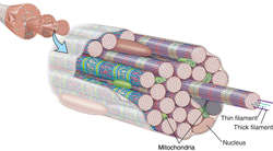
skeletal muscle
A tissue composed of muscle cells (often multinucleated) that contain neatly packed actin and myosin filaments; these filaments are arranged in cylindrical bundles called myofibrils. In each cell, the myofibrils are all aligned in the same direction and are parceled into longitudinal blocks (called sarcomeres) of similar lengths. Under the microscope, the ends of the blocks look like lines, making skeletal muscle cells appear to have regularly arranged striations. See: illustration
Skeletal muscle is innervated by somatic (as opposed to autonomic) motor axons at a synaptic structure called a motor endplate, where acetylcholine is the neurotransmitter. Most skeletal muscles can be controlled consciously, and skeletal muscle is sometimes referred to as voluntary muscle. Skeletal muscle cells contract more forcefully than smooth or cardiac muscle cells.
Skeletal muscle got its name because it usually attaches at one end to bone. Skeletal muscle is by far the most common type of muscle in the body and it plays a major role in normal metabolism, e.g., after a meal, excess glucose is removed from the blood stream primarily by skeletal muscle.
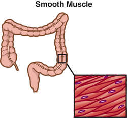
smooth muscle
A tissue composed of muscle cells that contain loosely-organized actin and myosin filaments. The lack of tight organization means that smooth muscle cells do not appear striated when examined under a microscope. Smooth muscle tissue tends to occur as sheets and is typically found in the walls of tubes, e.g., arteries, and sacs, e.g., the gastrointestinal system.
Smooth muscles are innervated by both sympathetic and parasympathetic autonomic motor axons; they are also stimulated by blood-borne molecules. Smooth muscles cannot be consciously controlled, and this form of muscle tissue is called involuntary muscle. Smooth muscle cells contract more slowly than skeletal or cardiac muscle cells.
soleus
Leg muscle. Origin: proximal ends of tibia and fibula. Insertion: calcaneus via Achilles tendon. Nerve: tibial (S1-S2). Action: plantarflexes foot.
See: leg for illus. (Muscles of the leg)somatic muscle
Muscle derived from mesodermal somites, including most skeletal muscle.
sphincter muscle
A muscle that encircles a duct, tube, or orifice, thus controlling its opening.
sphincter muscle of urinary bladder
The smooth muscle fibers around the origin of the urethra. Contraction of this muscle prevents urination; relaxation permits it.
stabilizer muscle
A muscle that supports a body segment so muscles attached to it can function.
stapedius muscle
Middle ear muscle. Origin: posterior wall of middle ear. Insertion: neck of stapes. Nerve: facial (CN VII). Action: tilts stapes, dampens excessive vibrations.
sternocleidomastoid muscle
Neck muscle. Origin: upper edge of manubrium, middle of upper clavicle. Insertion: mastoid process. Nerve: accessory (CN XI), spinal C2. Action: contralaterally rotates head.
See: face and headfor illus.striated muscle
See: tablesubscapularis muscle
Shoulder muscle. Origin: medial subscapular fossa. Insertion: lesser tubercle of humerus. Nerve: upper and lower subscapular (C5-C7). Action: medially rotates arm.
superior oblique muscle
Extraocular muscle. Origin: sphenoid bone deep in medial side of orbit. Insertion: lateral surface of eyeball behind its equator. Nerve: trochlear (CN IV). Action: turns eye down and outward with medial rotation.
superior rectus muscle
Extraocular muscle. Origin: tendinous ring around optic nerve at rear of orbit. Insertion: upper edge of eyeball in front of its equator. Nerve: oculomotor (CN III). Action: turns eye up and medially.
See: extraocular for illus.supraspinatus muscle
Shoulder muscle. Origin: medial supraspinous fossa of scapula. Insertion: greater tubercle of humerus. Nerve: suprascapular (C4-C6). Action: abducts arm.
synergistic muscles
Muscles aiding one another in function.
temporalis muscle
Muscle of mastication. Origin: temporal fossa of skull. Insertion: coronoid process of mandible. Nerve: trigeminal (CN V). Action: closes mouth, clenches teeth, retracts jaw.
See: headfor illus.tensor fascia lata muscle
Thigh muscle. Origin: iliac crest, anterior superior iliac spine. Insertion: iliotibial tract of fascia lata. Nerve: superior gluteal (L4-L5). Action: stabilizes (abducts) thigh, extends and laterally rotates leg.
tensor tympani muscle
Middle ear muscle. Origin: wall of auditory tube. Insertion: handle of malleus. Nerve: trigeminal (CN V). Action: tenses tympanic membrane, dampens excessive vibrations.
teres major muscle
Shoulder muscle. Origin: lower lateral edge of scapula. Insertion: bicipital groove of humerus. Nerve: lower scapular (C6-C7). Action: adducts and medially rotates arm.
teres minor muscle
Shoulder muscle. Origin: upper lateral edge of scapula. Insertion: greater tubercle of humerus. Nerve: axillary (C4-C6). Action: laterally rotates arm.
thenar muscle
The abductor or flexor muscle of the thumb.
thigh muscles
Anterior: iliopsoas, quadriceps (rectus femoris, vastus intermedius, vastus lateralis, and vastus medius), and sartorius muscles. Medial: adductor brevis, adductor longus, adductor magnus, gracilis, and pectineus muscles. Gluteal region: gemelli, gluteus maximus, gluteus medius, gluteus minimus, obturator externus, obturator, internus, piriformis, quadratus femoris, and tensor fasciae lata muscles. Posterior: biceps femoris, semimembranosus, and semitendinosus muscles.
See: leg for illus. (Muscles of the leg)thyroepiglottic muscle
A muscle arising on the inner surface of the thyroid cartilage. It extends upward and backward and is inserted on the epiglottis. It depresses the epiglottis.
tibialis anterior muscle
Leg muscle. Origin: lateral side of proximal tibia. Insertion: medial side of cuneiform bone, base of metatarsal 1. Nerve: deep peroneal (L4-L5). Action: inverts and dorsiflexes foot.
tibialis posterior muscle
Leg muscle. Origin: anterior tibia and fibula. Insertion: navicular, cuneiform, and cuboid bones; metatarsals 2-4. Nerve: tibial (L4-L5). Action: inverts and plantarflexes foot.
tonic muscle
Skeletal muscle fibers that contract slowly and that cannot propagate an action potential along their cell membranes. Tonic muscles are uncommon in humans and are found only in the extraocular muscles, stapedius muscle, and intrafusal fibers of the muscle spindles. The remainder of human skeletal muscle contains only twitch fibers.
trapezius muscle
Neck and back muscle. Origin: occipital bone (superior nuchal line), nuchal ligament, spinous processes of vertebrae C7-T12. Insertion: posterior edge of lateral clavicle, acromion, posterior edge of spine of scapula. Nerve: accessory (CN XI), spinal C3-C4. Action: elevates, retracts, and rotates scapula.
See: face and headfor illus.triangular muscle
A flat muscle with a broad origin and narrow insertion.
triceps muscle
Arm muscle. Origin: infraglenoid tubercle of scapula, posterior of proximal humerus, posterior of distal humerus. Insertion: olecranon process. Nerve: radial (C6-C8). Action: extends forearm.
Synonym: triceps brachii muscle See: arm for illus. (Muscles of the Arm)triceps brachii muscle
Triceps muscle.tricipital muscle
A muscle with three tendons of origin and a single, common insertion.
twitch muscle
Muscle fibers that can conduct axon potentials along their cell membranes. Almost all skeletal muscle in humans is twitch muscle. A very small number of muscles in humans are tonic muscles. Twitch muscles cells can be categorized into a number of types on the basis of the biochemical cycle that they use to produce their energy: red (oxidative), white (glycolytic), or intermediate (oxidative/glycolytic). Most human muscles are composed of a mix of twitch muscle cell types.
unipennate muscle
A muscle whose fibers converge on only one side of a tendon. See: bipennate muscle for illus.
unstriated muscle
Smooth muscle.uterine muscle
See: myometriumvastus intermedius muscle
Thigh muscle. Origin: anterior and lateral sides of proximal femur. Insertion: common tendon of quadratus muscles, tibial tuberosity via patellar ligament. Nerve: femoral (L2-L4). Action: extends leg.
vastus lateralis muscle
Thigh muscle. Origin: lateral side of proximal femur. Insertion: common tendon of quadratus muscles, tibial tuberosity via patellar ligament. Nerve: femoral (L2-L4). Action: extends leg.
See: leg for illus. (Muscles of the leg)vastus medialis muscle
Thigh muscle. Origin: medial side of femur Insertion: common tendon of quadratus muscles, tibial tuberosity via patellar ligament. Nerve: femoral (L2-L4). Action: extends leg.
vocalis muscle
Laryngeal muscle. Origin: midline of inner surface of thyroid cartilage. Insertion: arytenoid cartilage. Nerve: recurrent laryngeal of vagus (CN X). Action: changes tension of vocal cords.
voluntary muscle
A muscle that can be controlled voluntarily; most skeletal muscles are voluntary.
| Smooth | Cardiac | Striated | |
|---|---|---|---|
| Synonyms | Involuntary | Myocardial | Voluntary |
| Nonstriated | Skeletal | ||
| Visceral | |||
| Fibers | |||
| Length (in/m) | 50–200 | 25,000 | |
| Thickness (in/m) | 4–8 | 75 | |
| Shape | Spindles | Cylinders | |
| Markings | No striation | Striation | Marked striation |
| Nuclei | Single | Single | Multiple |
| Effects of cutting related nerve | Slight | Regulation of heart rate is lost | Complete paralysis |
extraocular muscle
Abbreviation: EOMSix muscles that attach outside the eyeball and that move the eye in its socket. The EOM are: the inferior and superior oblique muscles, and the lateral, medial, inferior, and superior rectus muscles.
See: extraocular for illus.Medical Dictionary, © 2009 Farlex and Partners
Source: https://medical-dictionary.thefreedictionary.com/EOM
Posted by: carterlithatinquir.blogspot.com
0 Response to "What Does Eom Stand for in Medical Terms"
Post a Comment